Purpose
To provide information on how to identify MEPs in EMG traces and how to determine MEP status. Simple MEP identification examples are provided.
Overview
A patient is classified as MEP+ if MEPs of any amplitude are observed in one or both muscles in 3 out of 10 traces at a given intensity. Only one MEP in either muscle is required at 100% MSO with bilateral facilitation to classify a patient as MEP+. Further details can be found in point 8 of the VERIFY TMS protocol.
A patient is classified as MEP- if MEPs cannot be elicited after attempting all of stimulation at 100% MSO while attempting voluntary facilitation and you have used systematic coil positioning to stimulate at least 5 different scalp locations using. All efforts need to be made to elicit a MEP if possible by using stimulation at 100% MSO, systematic coil positioning, and active bilateral facilitation.
Check the following before classifying a patient as MEP+:
MEP latency
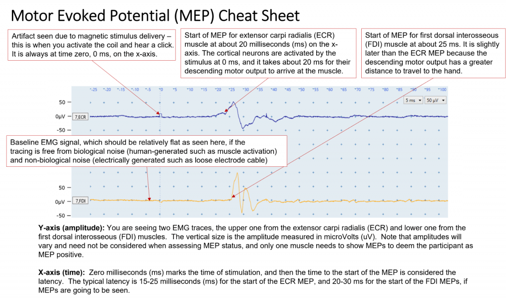
The latency of the MEP is the time between the cortical stimulation and the onset of the MEP in the target muscle. The onset of the MEP for ECR is typically 15 – 25 ms after the stimulus, and the onset for FDI is typically 20 – 30 ms after the stimulus. These latency windows account for the effects of stroke.
Latencies are also affected by the patient’s height. People who are below average height have latencies nearer the beginning of the window, and people who are above average height have latencies nearer the end of the window.
If MEPs are seen at appropriate latencies in either ECR or FDI then classify as MEP+.
If MEPs are only present in one muscle at a latency slightly later than expected then it is important to determine if a similar response is consistently seen in most EMG traces at approximately the same latency, within a couple of milliseconds. If this is the case, and the latency is only slightly outside the typical range then the patient may be classified as MEP+.
If responses are seen at random times across the patient’s EMG traces then this is indicative of spontaneous motor unit firing. Motor units firing can be identified due to their consistent size and shape in EMG traces. While the shape can sometimes resemble an MEP, they usually appear at latencies which do not correspond with the expected MEP latency. If you suspect motor unit responses rather than MEPs are present then a patient needs to be classified as MEP-, providing all efforts have been made to elicit a MEP if possible. Examples of EMG traces with motor units firing can be found in the EMG Technique module.
MEP consistency
Patients can be classified as MEP+ if you see MEPs in one or both muscles at consistent latencies in at least 3 out of 10 traces at a given stimulus intensity. Only 1 MEP in one or both muscles is needed to classify the patient as MEP+ if they are performing bilateral facilitation while you systematically move the TMS coil at 100% MSO. The amplitude of MEPs will likely vary between traces and this is not a factor when determining MEP status.
Examples of simple MEP identification
Here are some examples that classify patients as MEP+ or MEP- based on the presence of a MEP in one or both muscle traces at an appropriate latency.
Assume that each figure is representative of 3 EMG traces all collected with the same stimulus intensity at the ideal scalp location.
These examples were recorded using a MEGA-TMS TMS machine but the traces should look similar with different TMS units or EMG software.
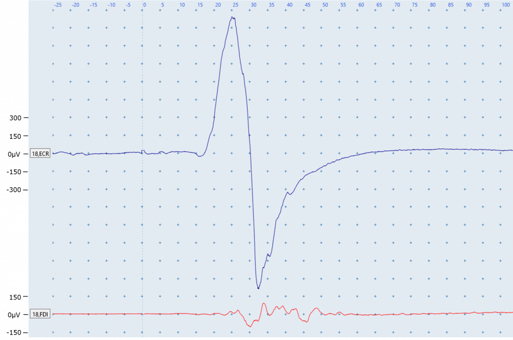
A: Patient (5ft 8”) at rest, 70% MSO
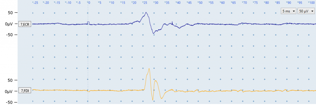
A: A MEP is identifiable in both the ECR (top trace) and FDI (bottom trace). The FDI MEP latency at 25 ms is slightly longer than ECR, as expected. This patient can be classified as MEP+.
B: Patient (5ft 4”) at rest, 90% MSO
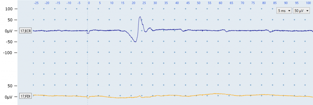
B: A MEP is identifiable at an appropriate latency of ~18 ms in the ECR EMG trace. No MEP is identified in the FDI trace. This patient can be considered MEP+, as MEPs in either ECR or FDI muscles allow a MEP+ classification.
C: Patient (6ft 0”) performing active facilitation, 100% MSO
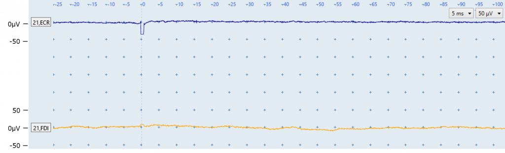
C: This patient had no MEPs at 100% MSO while at rest and is attempting active bilateral facilitation to increase the likelihood of eliciting a MEP. No MEPs are identified in either FDI or ECR EMG traces. The traces do not show muscle activity during facilitation due to severe paresis in the tested arm. If 4 other scalp locations had been stimulated prior to this figure also with no MEPs present then patient C can be classified as MEP-.
D: Patient (5ft 6”) at rest, 60% MSO
D: A MEP is identifiable in both ECR and FDI EMG traces. There is a clear large ECR MEP at an appropriate latency. The FDI MEP occurs at an appropriate latency and is much smaller than the ECR MEP, although note that MEP amplitude is not a determining factor for MEP status. The FDI MEP also demonstrates that MEPs don’t always have the stereotypical appearance that the ECR MEP has here. This patient can be classified as MEP+. It is worth noting that this patient would be considered MEP+ based on FDI MEPs alone, if ECR MEPs were not present.
Frequently asked questions
What should I do if there is a MEP in only one muscle, but the latency is a little later than I expect?
It is important to determine if a similar response is consistently seen in at least 3 of out 10 EMG traces at a latency that varies by no more than one or two milliseconds, using the same stimulator intensity. If this is the case, and the latency is only slightly outside the typical range then the patient may be classified as MEP+. If response latencies vary across traces this may indicate a random motor unit is firing, and that some responses are landing near the typical latency window by chance. Reviewing the shape of the response may help too, as responses due to motor unit firing tend to be very consistent in size and shape.
Quiz
Click here for a quiz to assess your learning on Simple MEP Identification.
Once the practice quiz has been completed with at least 70% correct you will be emailed a link to the final quiz for this module.
If you pass the final quiz with at least 80% correct you will be emailed a certificate of completion for the Simple MEP Identification module. There is no limit to the number of attempts for the practice or final quiz.
Please email verify.study.tms@gmail.com if you have any questions or comments.